Français
 Italiano
Größe dieser Vorschau: 724 × 600 Pixel. Weitere Auflösungen: 290 × 240 Pixel | 580 × 480 Pixel | 955 × 791 Pixel.
Originaldatei (955 × 791 Pixel, Dateigröße: 140 KB, MIME-Typ: image/jpeg)
Beschreibung
| Beschreibung | English: Chest X-ray of hyperinflated lung (9th anterior rib intersecting with the diaphragm. Normally, only the 6th rib will intersect with the diaphragm and the ribs below it will appear below the diaphragm) with bronchiectasis at the right hilum and right upper lobe of a 12 year old boy. Right hilum is at the same level as the left hilum which signifies right hilum elevation (left hilum is higher than the right hilum a normal chest X-ray). The most common cause of right hilum elevation is right upper lobe collapse (not seen in this study). Reverse "7" or "7" sign of the bilateral ribs which signifies pectus excavatum. When horizontal posterior ribs meets with the anterior ribs, the resultant shape is similar to a "7" on the left lung, and mirror image of "7" in the right lung. A normal chest X-ray usually shows posterior rib curves downwards to meet with the anterior rib. Thus, this patient may have bronchiolitis obliterans with underlying pectus excavatum. |
| Datum | |
| Quelle | Eigenes Werk |
| Urheber | Cerevisae |
Lizenz
| This file is ineligible for copyright and therefore in the public domain, because it is a technical image created as part of a standard medical diagnostic procedure. No creative element rising above the threshold of originality was involved in its production. See Meta:Wikilegal/Copyright of Medical Imaging for details.
|  |
Dateiversionen
Klicke auf einen Zeitpunkt, um diese Version zu laden.
| Version vom | Vorschaubild | Maße | Benutzer | Kommentar | |
|---|---|---|---|---|---|
| aktuell | 16:06, 16. Jan. 2022 | | 955 × 791 (140 KB) | Cerevisae | Uploaded own work with UploadWizard |
Dateiverwendung
Keine Seiten verwenden diese Datei.
Metadaten
Explore the world
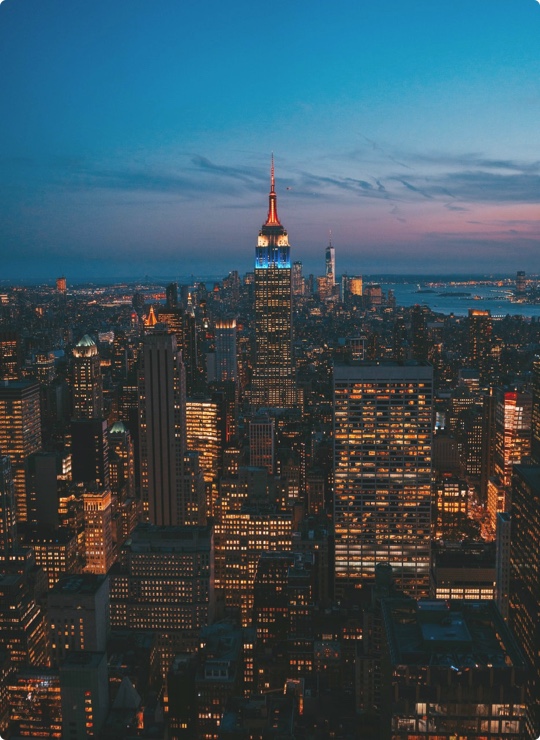
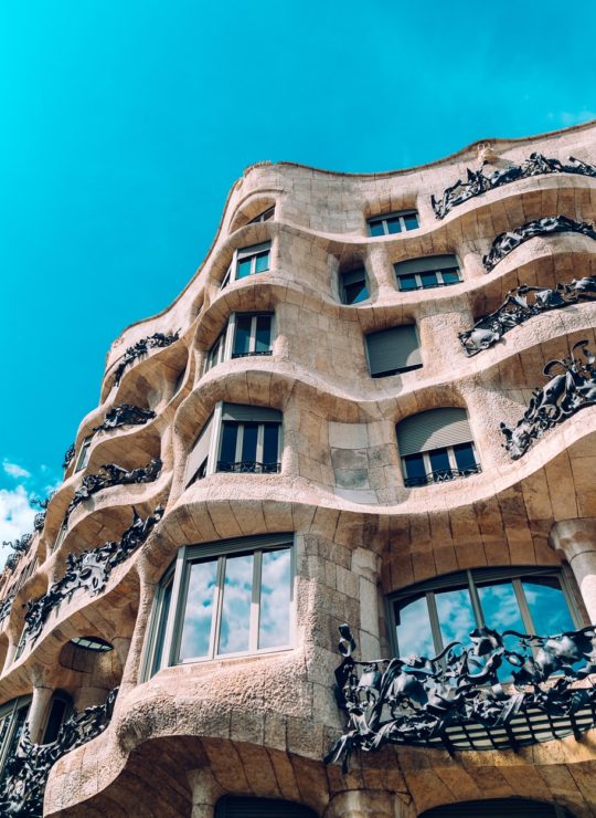
United states
New York
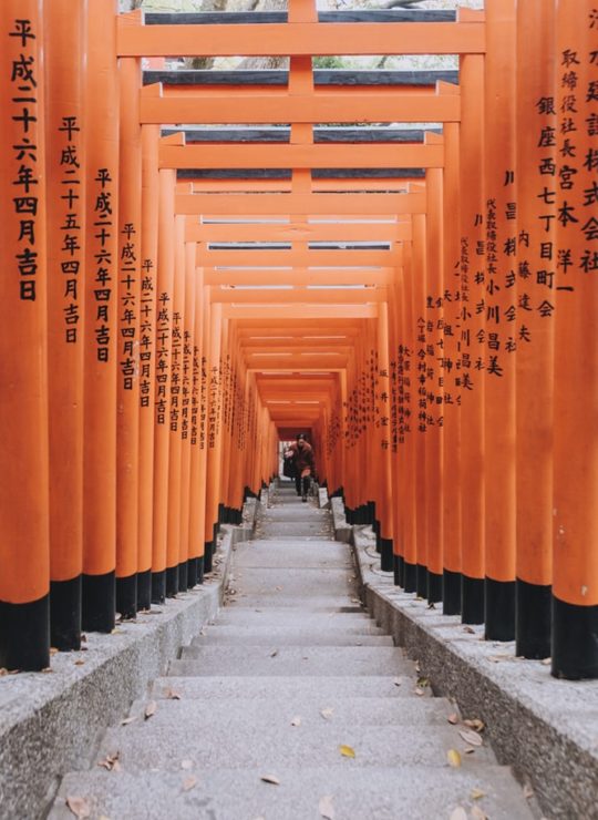
Japan
Tokyo
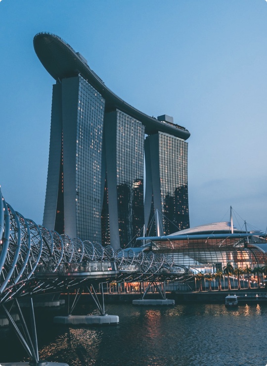
Singapore
Singapore